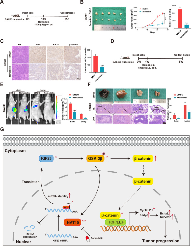
Fig. 8.
The role of remodelin in vivo and a schematic model for the mechanisms of NAT10. A The schematic diagram of the application of remodelin in the xenograft models of mice. B Representative images of subcutaneous xenograft tumors (n = 5 for each group). The tumor volumes were measured every 5 days and the tumor weights were analyzed. C HE and IHC staining of xenograft tumors. The expression of Ki67, KIF23, and β-catenin were detected by IHC. D The schematic diagram of the application of remodelin in the metastasis models of mice. E Representative images and analysis of luminescence intensity in metastasis models (n = 5 for each group). F Representative image and HE staining of metastatic tumors in the livers and lungs of mice. The number of metastases in livers or lungs was analyzed. G The schematic model for the mechanisms of NAT10 in CRC. All data are presented as mean ± SD. **P < 0.01, ***P < 0.001, ****P < 0.0001

