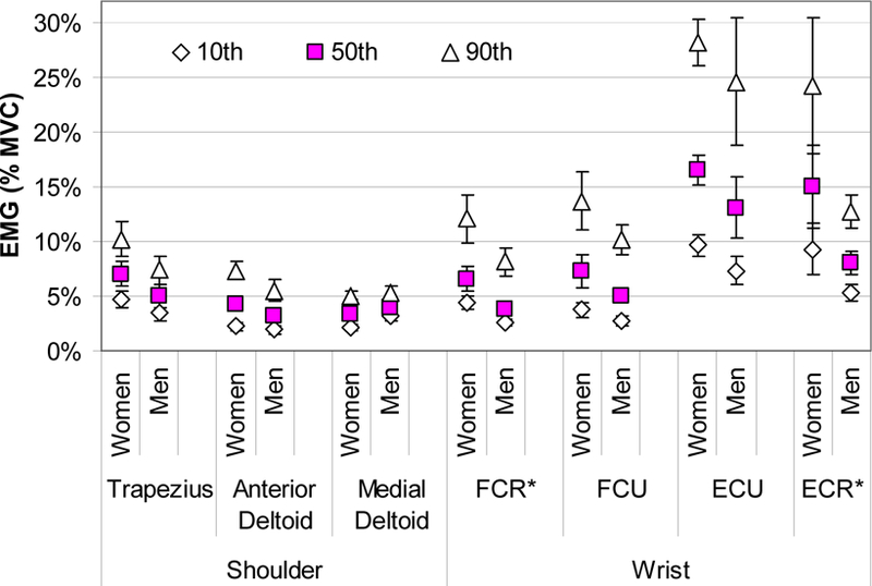
Figure 1.

Average shoulder and forearm EMG amplitudes (10th (◊), 50th (□) and 90th (∆) percentile) grouped by gender for the standard workstation configuration. The error bars represent the standard error across subjects within the gender group. Females tended to have higher muscle activity in all but the medial deltoid muscle. Student t-tests compared the values between the subjects grouped by gender with significance found for the 10th and 50th percentile for FCR and the 50th and 90th percentile for the ECR.
