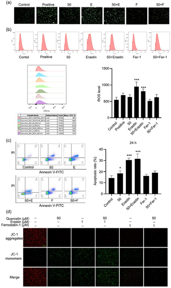
Fig. 4.
Quercetin induced HEC-1-A cell apoptosis by regulating ferroptosis. a, b ROS level in HEC-1-A cells. Cells were treated with quercetin for 24 h, then the ROS level was detected by DCFH-DA staining and cells were visualized under a fluorescent microscope and analyzed by flow cytometry. c Cell apoptosis detection by flow cytometry. Cells were treated with quercetin for 24 h, and the apoptotic cells were stained with Annexin V-FITC/PI followed by flow cytometry. d Detection of mitochondrial membrane potential of HEC-1-A cells by JC-1 staining. Cells were treated with quercetin for 24 h, and visualized under a fluorescent microscope. Red fluorescence represents the mitochondrial aggregate JC-1 and green fluorescence indicates the monomeric JC-1. Data are expressed as mean ± SD (n = 3). *p < 0.05, ***p < 0.001 vs. Control

