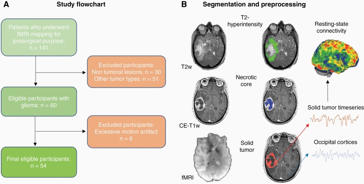
Figure 1.
Study flowchart and MRI preprocessing. (A) A retrospective analysis was conducted on all patients who underwent rs-fMRI at BWH (Boston, MA, US) between September 1, 2012 and September 1, 2018 because of a suspected brain focal lesion. (B) Gliomas were manually segmented into their necrotic core/surgical cavity, solid region (mass) and T2-hyperintense area on the basis of T1w and T2w MRI scans. Average BOLD timeseries were extracted from the solid tumor mask of each patient and correlated with the rest of the brain at single voxel-level, using age and gender as covariates. CE-T1w, contrast-enhanced T1w; fMRI, functional Magnetic Resonance Imaging.

