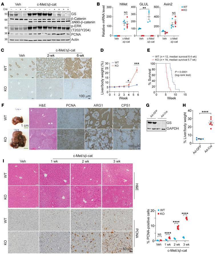
Figure 1. Hepatic ablation of GS exacerbates HCC development driven by c-Met/ΔN90-β-catenin.
Seven-week-old Glulfl/fl Alb-Cre– (WT) and Glulfl/fl Alb-Cre+ (KO) male mice were injected with either vehicle or c-Met/ΔN90-β-catenin/SB10 plasmids via SB-HTVI. Livers were collected and analyzed at the indicated time intervals. (A) Immunoblotting of liver tissue samples collected at 6 weeks (endpoint of the KO mice) after HTVI. Representative blots are shown (n = 3–5). The protein molecular weight in kDa is indicated on the left. (B) Relative mRNA levels in livers 2 weeks after HTVI were determined by qPCR (n = 3–4). (C) IHC of GS was performed at 0, 2, or 6 weeks after HTVI (n = 3). Representative images are shown. (D) Liver/body weight ratios were compared (n = 3–6). (E) Kaplan-Meier curves are shown. (F) Representative gross, H&E, and IHC images of liver tissues harvested 6 weeks after oncogene injection (n = 3). (G and H) Seven-week-old Glulfl/fl male mice were first coinjected with pCMV-c-Met/ΔN90-β-catenin plasmids. One week later, half of the mice were randomly selected and injected with adenoviral CMV-Cre (Ad-Cre), while the other half were injected with adenoviral CMV-GFP (Ad-GFP) as controls via the tail vein. Livers were harvested another 7 days later, and immunoblotting showed successful GS knockout by Ad-Cre (G). Mice were harvested at the endpoint (6 weeks after HTVI injection) (n = 6). Liver weight/body weight ratios were compared. The results are expressed as mean ± SEM (H). (I) Liver sections from WT and KO mice were obtained at the indicated time points and processed for H&E and PCNA IHC staining (n = 3 mice for each group). Representative images are shown. The number of PCNA-positive cells was quantified by ImageJ from 6 randomly selected fields. Shown on the right is the mean percentage ± SEM. *P < 0.05; **P < 0.01; ***P < 0.001; ****P < 0.0001 by 2-tailed t test (B, D, and I). NS, not significant. Scale bars: 100 μm (C and F [right]), 1 cm (F, left), and 50 μm (I).

