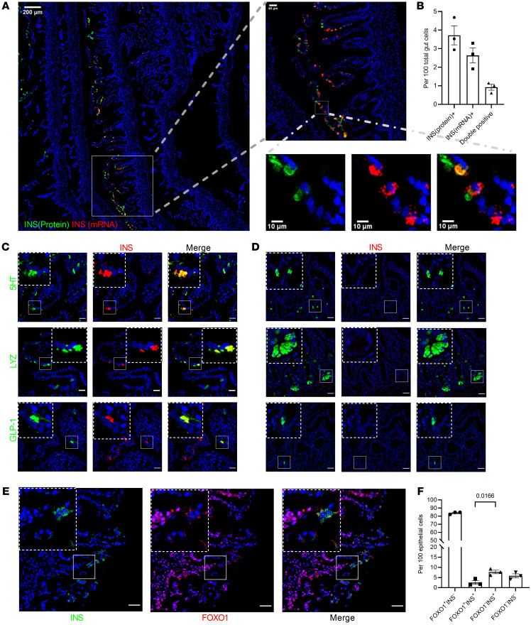
Figure 1. INSULIN and FOXO1 expression in human fetal small intestine secretory lineage cells.
(A) Representative image (GA = 17 weeks) of tile scanning of one-fourth fetal proximal intestinal roll section stained with INS mRNA in red and INS protein in green. (B) Quantification of INS protein+, INS mRNA+, and double-positive cells. n = 3 different donors. GA = 15–17 weeks. Data are represented as mean ± SEM. (C) Insulin (red) and 5HT, lysozyme, or GLP-1 (green) staining in fetal human anterior intestine (GA = 17 weeks). Colocalization is shown in yellow. Scale bars: 20 μm. (D) Insulin (red) and 5HT, lysozyme, or GLP-1 (green) staining in adult human duodenum. Colocalization is shown in yellow. Scale bars: 40μm. (E) Insulin (green) and FOXO1 (red) staining in fetal human anterior intestine. Scale bars: 20 μm. (F) Quantification of FOXO1–Insulin+ versus FOXO1+Insulin+ cells in fetal human proximal intestine. n = 3 different donors. Each point shows averaged counting value from 3 to 4 different images per donor. Data are represented as mean ± SEM. Two-tailed t test.

