. 2022 Dec 5;11:e78609. doi: 10.7554/eLife.78609

Figure 4. Glycine targets NINJ1 oligomerization to prevent membrane rupture in primary human macrophages.

(A) Native-PAGE analysis of endogenous NINJ1 from human monocyte-derived macrophages (hMDMs) stimulated to undergo pyroptosis displays a shift to higher molecular weight, which is abrogated by glycine treatment. NINJ1 levels are almost absent in siNINJ1-treated cells (quantification in Figure 4—figure supplement 2A), and no shift is seen after LPS and nigericin treatment. (B) Total internal reflection fluorescence (TIRF) microscopy of LPS-primed primary hMDMs shows endogenous NINJ1 in discrete plasma membrane puncta. Cell membrane outline (dotted white line) was determined using fluorescently labeled cholera toxin subunit B (not shown) as a plasma membrane marker. Scale bar 20 μm. Anti-human NINJ1 antibody validation for immunofluorescence is provided in Figure 4—figure supplement 1. Quantification of the NINJ1 puncta density (C) and fluorescence intensity (D) in LPS-primed hMDMs at baseline or stimulated to undergo pyroptosis (nigericin 20.7 μM for 2 hr) without or with glycine (50 mM). Violin plot of NINJ1 puncta density and intensity quantified from images of cells from four independent donors. Mean of NINJ1 puncta densities from three cells per donor per condition shown as superimposed datapoints (different symbols are used for individual donors) along with median and quartiles for each condition.

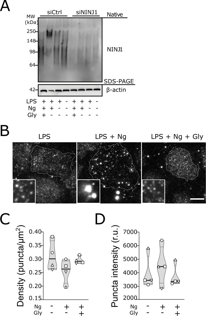
Figure 4—source data 1. Numerical values for experimental data plotted in Figure 4.
elife-78609-fig4-data1.xlsx (337.9KB, xlsx)

Figure 4.

Figure 4—figure supplement 1. Mouse anti-human NINJ1 monoclonal antibody validation.

Figure 4—figure supplement 1.

LPS and nigericin treated human monocyte-derived macrophages (hMDMs) were fixed and stained with the indicated antibodies as described in the main text prior to imaging by total internal reflection fluorescence (TIRF) microscopy. (A) Cells were stained with mouse anti-human NINJ1 (IgG2b) Ab or a control Ab (mouse IgG2a) followed by fluorescent rabbit anti-mouse Ab. The cell plasma membrane was stained using fluorescent cholera toxin subunit B (CTxB). (B) In a separate experiment, cells were stained with mouse anti-human NINJ1 (IgG2b) Ab or no primary Ab, followed by fluorescent rabbit anti-mouse Ab. Scale bars 20 μm.
Figure 4—figure supplement 2. Glycine prevents NINJ1 aggregation and pyroptotic lysis in induced pluripotent stem-cell (iPSC) derived human macrophages (iPSDMs) without affecting IL-1β secretion.

Figure 4—figure supplement 2.

(A) Quantification of NINJ1 native-PAGE signal from unstimulated human monocyte-derived macrophage (hMDM) lysates confirm NINJ1 silencing in siNINJ1-treated cells compared to siCtrl-treated cells. (B–D) iPSDMs were LPS-primed and stimulated to undergo pyroptosis with nigericin in the presence or absence of 50 mM glycine. (B) Native-PAGE analysis of endogenous NINJ1 in iPSDMs display a shift to higher molecular weight when cells are stimulated to undergo pyroptosis, which is abrogated by glycine pretreatment. (C) LDH release was measured in the supernatants to assess cell rupture and was reduced with glycine co-treatment. Data are expressed as % of LDH in supernatant from LPS and nigericin-treated cells in n=3 independent experiments. Data points from each experiment is shown along with their mean and SD. (D) IL-1β release from pyroptotic cells was not altered by the presence of glycine. Data points from three technical replicates from one experiment are shown along with their mean and SD.
Figure 4—figure supplement 2—source data 1. Numerical values for experimental data plotted in Figure 4—figure supplement 2.