. 2022 Oct 27;11:e83373. doi: 10.7554/eLife.83373

Figure 5. Bolus dosing with different LDEs or reactive covalent electrophilic drugs elicits complex AR responses.

Also see Figure 6, Figure 3—figure supplements 1 and 4 and 5E, Figure 4—figure supplement 1, and Figure 5—figure supplement 1. (A) Representative IF images of Tg(gstp1:GFP) fish expressing Halo-•-hKeap1-(2xHA), following bolus exposure to indicated electrophiles. (B) Quantitation of data in (A). Image quantitation was performed on the head/tail-regions as illustrated. No. embryos analyzed: Tail, DMSO (55), Sulforaphane (16), HNE (12), Tecfidera (24), NE (9); Head, DMSO (43), Sulforaphane (16), HNE (15), Tecfidera (24), NE (9). Sulforaphane and HNE elicit non-significant and 1.5-fold AR upregulation, respectively. Tecfidera gives medium (~2-fold) AR response and NE elicits the strongest AR upregulation (~3-fold) in tail. Consistent with data elsewhere (e.g., Figure 3A and C-E), head is not responsive. (C) qRT-PCR analysis of endogenous AR-responsive genes following bolus exposure of native reactive LDEs to whole fish similarly shows mixed responses. Whole-head/-tail separation was performed as indicated in inset (left), prior to RNA isolation selectively from the tails. 2 hr post Z-REX, embryos were euthanized, and RNA was isolated separately from tail (see inset, left). Data are presented as mean ± sem. n>3 independent biological replicates and two technical repeats for each sample. All numerical data present mean ± sem. Numbers above the bars represent analysis by two-tailed t-tests.

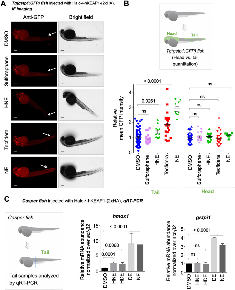
Figure 5—source data 1. Quantification results.

Figure 5.

Figure 5—figure supplement 1. Uncontrolled bulk exposure of developing larvae with reactive electrophiles results in differential proteome labeling extent and adversely affects development/viability.

Figure 5—figure supplement 1.

(A) Treatment with 25 µM CDDO-Me for 4 hr negatively impacts the morphology of the developing embryos (curved tail; malformed/missing caudal fin), whereas Z-REX photocaged probe treatment shows no negative impacts. See also Figure 3—figure supplement 1. Top: CDDO-Me treated, Bottom: Ht-PreNE treated. (B) Comparison of relative whole-reactive-proteome labeling extent by the respective alkyne-bearing small-molecules, following either bolus LDE treatment (2nd to 4th bars) or treatment with Z-REX-photocaged-LDE precursor alone (6th bar) or full Z-REX execution (7th bar) in live embryos. Zebrafish embryos at 32 hpf were directly treated with 25 μM of the indicated LDEs for 2 hr. After fixing and permeabilization, Cy5 azide was conjugated to alkyne labeled proteins using copper-assisted Click procedure as described in Methods. Larvae were imaged after washing away unbound dye. The Cy5 fluorescence intensity was analyzed using Image-J(NIH). Electrophiles devoid of 4-OH group such as DE and NE (4th and 5th bars) (see also Figure 1A inset) efficiently label the whole-reactive-proteome upon bolus treatment of whole embryos, whereas 4-OH-group containing electrophile such as HNE and HDE (2nd and 3rd bars) show a lower degree of whole-reactive-proteome labeling. See Figure 6 for representative images related to the data in 1st to 5th bars. All numerical data present mean ± sem. Numbers above the bars represent analysis by two-tailed t-tests.
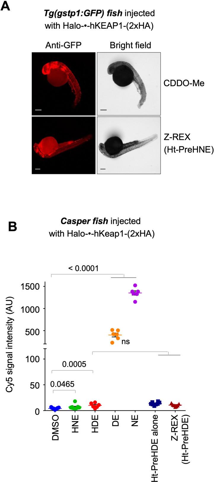
Figure 5—figure supplement 1—source data 1. Quantification results.