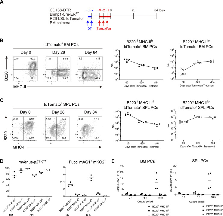
Figure 2.
Differentiation of B220loMHC-IIlo PCs from B220hiMHC-IIhi PCs. (A and B) BM chimeric mice were generated by transfer of BM cells from CD138-DTR Blimp1-CreERT2 R26-LSL-tdTomato mice into x-ray irradiated mice. In the chimeric mice, pre-existing PCs derived from donor BM cells were depleted by DT treatment (Fig. S3, A–C), and de novo–generated PCs from the same BM cell origin were subsequently labeled by tamoxifen administration. These tdTomato+ PCs were analyzed by FCM at the indicated time points. (A) Schematic of the experimental procedure. (B and C) The expression of B220 and MHC-II on tdTomato+ PCs and the absolute numbers of B220hiMHC-IIhi and B220loMHC-IIlo tdTomato+ PCs in BM (B) and SPL (C) at the indicated time points after tamoxifen treatment. Four or five mice were analyzed at the indicated time points. (D) Cell cycle status of each PC subset from SPL or BM analyzed by using mVenus-p27K- mice (left, G0) and Fucci mice (right, S, G2, M). Three mice each were analyzed. (E) The frequency of late apoptotic (CaspGLOW+ PI+) cells in each PC subset after in vitro culture for the indicated times. PCs isolated from four mice were analyzed. Data are representative of two independent experiments.

