Figure 5.
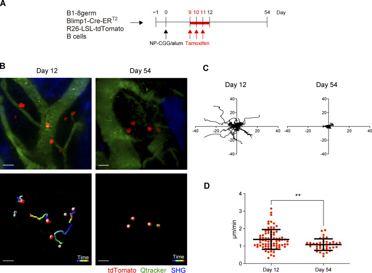
Maturation-dependent PC dynamics in the BM niche. B6 mice were transferred with splenic B cells from B1-8germ Blimp1-CreERT2 R26-LSL-tdTomato mice, immunized with NP-CGG in alum, and then treated with tamoxifen from day 9 to 11 after immunization. Intravital BM imaging was performed using these mice on day 12 and 54 after immunization. (A) Schematic of the experimental procedure. (B) A representative maximum-intensity projection image of skull bone tissues from the mice at the indicated time points after immunization (top). Red, PCs expressing tdTomato; green, blood vessels (Qdot); blue, bone tissues (SHG). Scale bar, 20 μm. Trajectory of these PCs measured for 1 h (bottom). White spheres were automatically generated by Imaris software recognizing tdTomato+ cells. (C) Displacement of PCs in the x–y plane during 1 h of observation in the mice at the indicated time points after immunization. (D) Mean track speed of PCs at the indicated time points. Data are pooled from two mice with 77 cells and three mice with 37 cells at day 12 and 54 after immunization, respectively. All data are pooled from two independent experiments. **P < 0.01 by two-tailed unpaired Student’s T test. The imaging data are available in Video 1.

