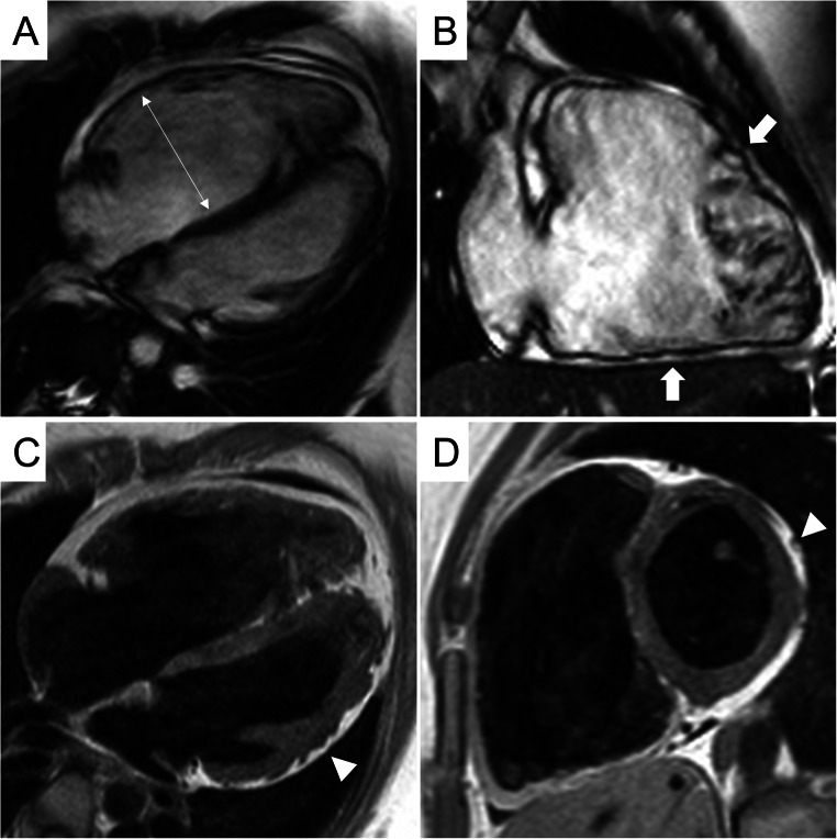
Fig. 2.
CMR frames of a patients with biventricular ACM. Morpho-functional abnormalities of the RV can be appreciated on 4-chamber view (A) and right ventricular 2-chamber long-axis view (B) of cine images, evidencing RV dilatation (A, double-head arrow) and multiple sacculations of the inferior and RVOT regions (B, arrows). Structural alterations of the LV are showed in 4-chamber view (C, arrowhead) and short-axis view (D, arrowhead) of T1-weigheted images, where fibro-fatty infiltration of the infero-lateral LV walls becomes evident as a hyperintense signal with a typical bite-like pattern. ACM, arrhythmogenic cardiomyopathy; LV, left ventricle; RV, right ventricle; RV, right ventricle outflow tract

