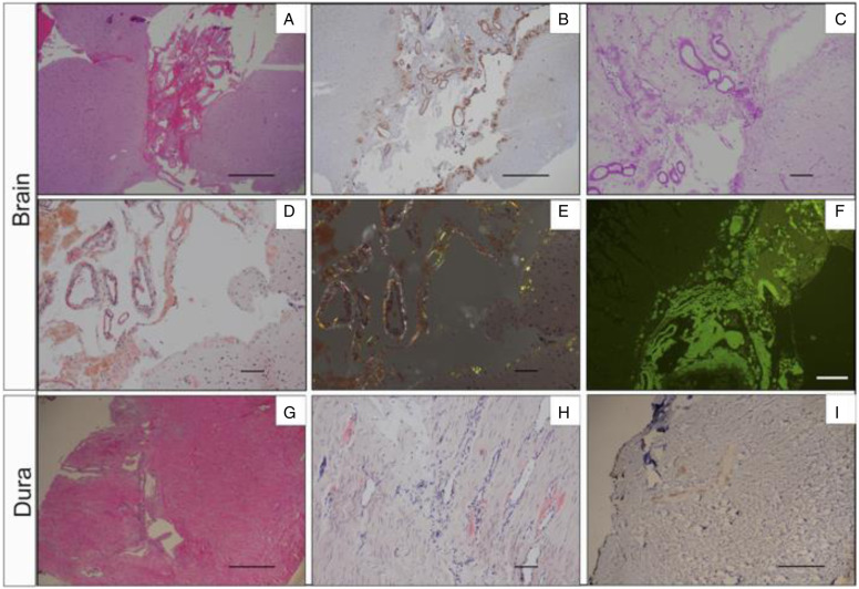
Figure 3.
Brain and dura biopsy. (A-F) Right temporal brain biopsy shows transthyrentin deposition in both leptomeninges and subpial brain parenchyma. (A). H&E image; (B). Transthyrentin (prealbumin) immunostaining; (C). PAS stain; (D). Congo red special strain; (E). Apple-green birefringence with polarized light; (F). Thioflavin immunofluorescence stain. (G-I) Right temporal dura biopsy displays transthyretin deposition in blod vessel walls. (G). H&E image; (H). Congo red special stain; (I). Beta-amyloid immunostaining. A, B, G, H, I: scale bar = 500μm; C, D, E, F: scale bar = 100μm.

