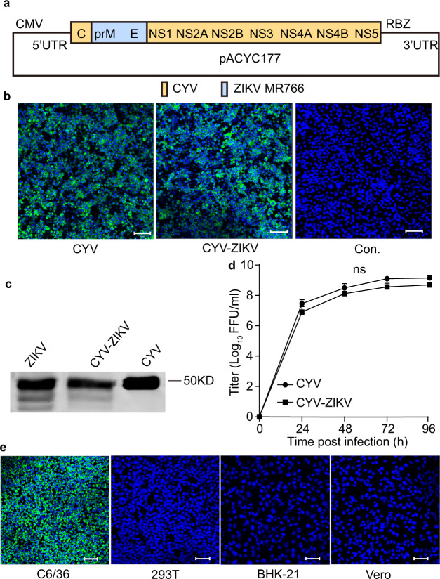
Fig. 1. Characterization of the CYV-ZIKV chimeric virus.
a Schematic diagram of the CYV-ZIKV chimeric virus infectious clone. The encoding sequence of the prME of the CYV (yellow) was replaced by that of the ZIKV MR766 strain (blue). The viral genome was placed between a CMV promoter and a RBZ in the pACYC177 vector. b The wild-type CYV and CYV-ZIKV chimeric viruses were rescued by transfection of the viral infectious clone plasmids into C6/36 cells. ZIKV E protein expression in C6/36 cells was detected by immunofluorescence with mAb 4G2 (green), and the nucleus was stained with Hoechst 33342 (blue) 3 days post-transfection. Controls (Con.) was not transfected. The bars indicate 50 μm. c C6/36 cells were infected with CYV, ZIKV, or CYV-ZIKV at a MOI of 1. The virus supernatants were purified by sucrose gradient centrifugation at 3 dpi, separated by western blot, and stained by anti-E mAb 4G2. The source data was provided in Fig. S10. d Growth curves of CYV and CYV-ZIKV after infection of C6/36 cells at a MOI of 1. The supernatant titers were gauged with a focus-forming assay on C6/36 cells (n = 3). Data are presented as means ± standard deviation (SD) of triplicate measurements. The P-value was determined by a two-sided multiple t-test, and ns indicates not significant. e Susceptibility of CYV-ZIKV on mosquito and vertebrate cells. The cells were infected with CYV-ZIKV at a MOI of 0.1. ZIKV E protein expression in C6/36 cells was detected by immunofluorescence with mAb 4G2 (green), and the nucleus was stained with Hoechst 33342 (blue) at 3 dpi. C6/36 (A. albopictus cell line), 293T (human embryonic kidney cell line), BHK-21 (Baby hamster kidney cell line), and Vero (African green monkey kidney cell line). The bars indicate 50 μm. Similar results were obtained in three independent experiments. Source data are provided as a Source Data file.

