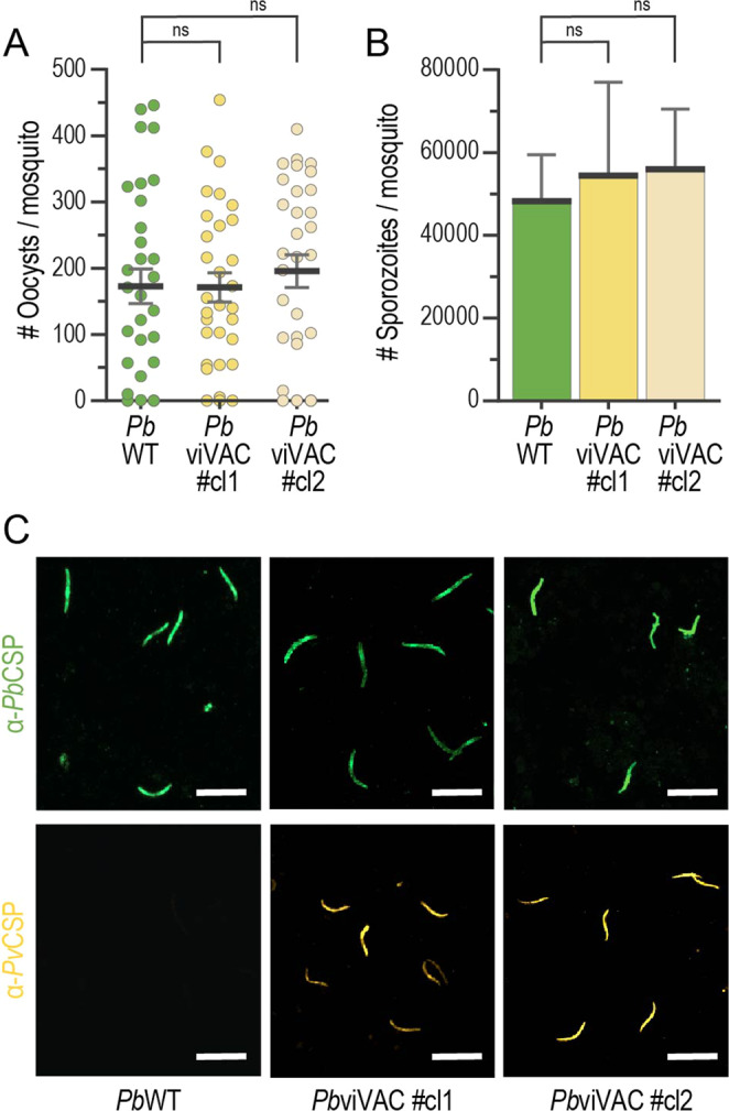
Fig. 2. PbviVac sporogonic development and PvCSP expression by salivary gland sporozoites.

A, B Midgut oocyst and salivary gland sporozoite numbers in PbWT and PbviVac-infected mosquitoes (n ≥ 28 mosquitoes per group); C Representative immunofluorescence microscopy images of PbCSP (green) and PvCSP (yellow) expressed by PbWT (left) and PbviVac (middle and right) sporozoites. Scale bar: 20 µm. Measurements were taken from distinct samples. The black lines/bars and grey lines correspond to mean and standard error of the mean, respectively (ns: not significant, Mann–Whitney U test).
