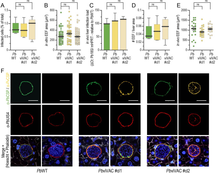
Fig. 3. PbviVac in vitro and in vivo pre-erythrocytic development and expression of PvCSP.
A, B Compared in vitro infectivity and parasite development of PbWT and PbviVac parasites in HepG2 human hepatoma cells (n ≥ 3 coverslips per group); Compared in vivo infectivity and development of PbWT and PbviVac parasites as determined by qPCR analysis of infected mouse livers (C), quantification of the number of parasites per liver area (D), and development of hepatic parasites (E) (n = 3 mice per group); (F) Representative immunofluorescence microscopy images of PbWT and PbviVac parasites developing in mouse livers 48 hpi. Immunofluorescence staining with the anti-PbCSP (green) and anti-PvCSP VK210 (yellow), as well as with anti-PbUIS4 antibodies, confirms the expression of both proteins by PbviVac and their localization to the parasite membrane. Scale bar: 20 µm. Measurements were taken from distinct samples. The boxes correspond to the 25th and 75th percentiles in (A) and (D) and the black lines/bars and grey lines correspond to mean and standard error of the mean, respectively (ns: not significant, Mann–Whitney U test).

