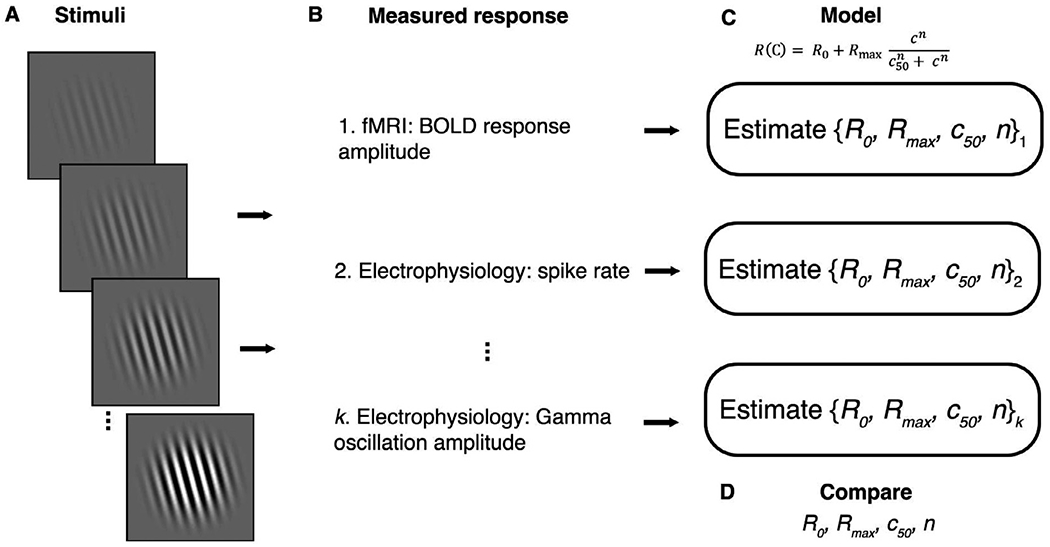
Fig. 1.

The neural basis of the fMRI signal can be tested by comparing responses with reference to parametric variations of an input stimulus. (A) A set of stimuli are chosen that parametrically vary in some dimension - in this case, spatial contrast. (B) Measurements are made in multiple modalities. These measurements do not need to be made simultaneously or even in the same individuals or species. (C) Responses are modeled as a function of the stimulus using the same model form (but different fitted parameters) for each modality. For example, one can estimate R0, Rmax, c50, and n for different measurement types in response to variations in stimulus contrast. (D) The model parameters are compared between multiple measurement types with reference to parametric variations in stimulus contrast. Note that the measurements in (B) are not directly compared to each other.
