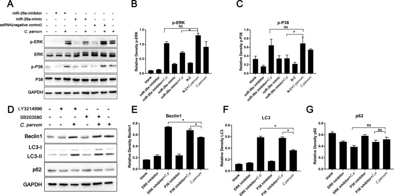
Fig. 5.
MiR-26a-induced autophagy in HCT-8 cells infected with C. parvum via p38 and ERK signaling. HCT-8 cells were transfected with miR-26a-mimic, miR-26a-inhibitor or ssRNA (negative control) and were infected with C. parvum sporozoites at 24 h post-transfection. Protein samples were collected at 12 h post infection and were analyzed by western blotting (A). The results were further analyzed by grayscale analysis from three independent experiments (B, C). HCT-8 cells were treated with ERK inhibitor LY3214996 (1 μM) and P38 inhibitor SB203580 (5 μM) for 1 h and then were infected with C. parvum sporozoites. The protein samples were collected at 12 h post infection and were detected by western blotting (D). The results were further analyzed by grayscale analysis from three independent experiments (E–G). A one-way ANOVA assay with Tukey-Kramer post hoc test was used for analyzing data. Data are expressed as the mean ± SD from three independent experiments (*P < 0.05, ns = no significant differences)

