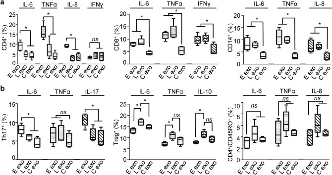
Figure 3.
Cytokine production in subsets of immune cells in response to COVID-19 plasma exosomes. (a) MicroBeads sorted CD4+ T cells, CD8+ T cells, and CD14+ monocytes were treated with plasma exosomes (4 × 10⁹ ml−1) from the same patients (average age 52.1) early (E exo) in their admission and later (L exo) in their hospitalization (average 4 days) or non-COVID-19 donors (C exo) at 37 °C for 16 h. Cytokine production was quantified using flow cytometry gated for live CD4+, CD8+, CD14+ cells. (b) Cytokine production was quantified using MicroBeads selected CD4+ cells gating on Th17 T cells using PE-CF594-conjugated CD196 (CCR6) (Clone 11A9, BD Bio.) and on Treg T cells using PE-conjugated CD25 (Clone 2A3, BD Bio.). CD4+ central memory T cells were separated using MicroBeads kit (Miltenyi Biotech) and gated for live cells. Isotype controls and no-antibody blank controls were used in parallel in flow cytometry. Data represent average ± SD; n = 10; *p < 0.05; ns, p > 0.05; one-way ANOVA equal variant.

