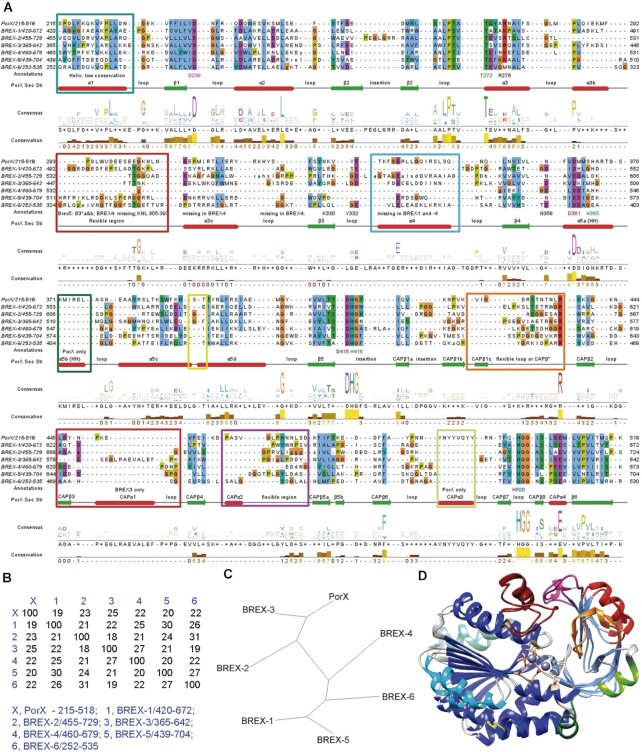
Figure 9.
Comparison of the primary and tertiary structures of the PglZ domains in PorX and the BREX1–6 PglZ proteins. (A) Multiple sequence alignment of the PorX PglZ domain with all PglZ domains of BREX-1 to BREX-6. (B) Percent identity matrix and (C) the phylogenetic tree from the primary sequence alignment. The BREX PglZ proteins are a highly divergent group with sequence identities within the PglZ domain of 19–30%, but the conserved fold predicted by AlphaFold 2 modelling nevertheless closely resembles the PglZ domain of PorX (Supplementary Figure S10). The BREX-3 PglZ shows the highest similarity to the PorX PglZ domain, with an identity of 25%. (D) Superposition of PorX-PglZ and BREX-3 PglZ. Regions with major differences are marked in colour in the structural superposition and as coloured rectangles in the alignment. The shared PglZ folds catalytic subdomain are coloured in blue and the cap subdomains are coloured in light blue.

