FIG. 2.
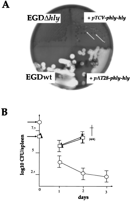
Complementation. (A) In vitro complementation. The hemolytic phenotype of L. monocytogenes colonies was visualized on horse blood agar plates after 24 h of incubation at 37°C. The two strains denoted + pTCV-pphly-hly and + pAT28-phly-hly correspond to EGDΔhly transformed with the corresponding plasmids. The arrows show the two types of colonies (Hly+ and Hly−) observed with the strain transformed with pTCV-phly-hly in the absence of antibiotic selection (Hly− is indicated by a star). (B) In vivo complementation. The kinetics of infection was followed in the spleens of infected mice. With the negative control, EGDΔhly (○), mice were inoculated with 2 × 10⁹ bacteria/mouse (indicated by an arrow to the left of the ordinate); with the two transformed derivatives, mice were inoculated with 2 × 10⁷ bacteria/mouse (arrow) (□, EGDΔhly transformed with pTCV-phly-hly; ▵, EGDΔhly transformed with pAT28-phly-hly). †, death; four mice out four infected died.

