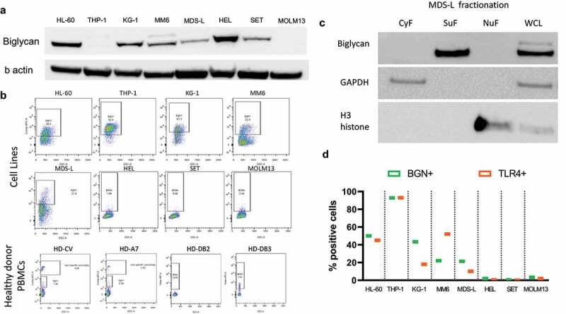
Figure 1.

Expression and localization of BGN in AML/MDS cell lines. (a) Western blot analysis was performed as described in Materials and Methods using an anti-BGN-specific Ab. Staining with an anti-GAPDH Ab served as loading control. A differential BGN expression was found in the 4 AML (HL-60, THP-1, KG-1, MM6), 2 MPN (HEL, SET) one MDS (MDS-L) and one sAML (MOLM13) cell lines. (b) Dot plots revealing the surface expression of BGN in the 8 cell lines as well as PBMCs of four healthy donors. (c) Spatial distribution of BGN in the MDS-L cell line after cell fractionation. Three fractions are investigated (cytoplasmic fraction CyF, surface fraction SuF, nuclear fraction and cytoskeleton NuF) along with a whole cell lysate as a control. Successful separation of fractions was determined using an anti-GAPDH Ab and an anti-H3 histone Ab for the CyF and NuF fractions, respectively. (d) Comparison of the surface expression of BGN (green) and TLR4 (Orange) on the 8 cell lines. The cells were stained in separate samples for both BGN and TLR4 as described in Materials and Methods and their expression was compared afterward.
