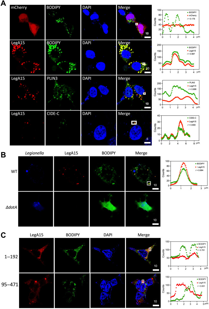
Fig. 1. LegA15 targets host LDs.
(A) Ectopically expressed LegA15-mCherry colocalizes with host LDs. Biomarkers PLIN3 and CIDE-C were shown in red by their antibodies. (B) LegA15 secreted by L. pneumophila colocalizes with the host LDs. The L. pneumophila WT and ΔdotA strains carrying a complemented LegA15 plasmid infected macrophage bone marrow–derived macrophage (BMDM) cells for 2 hours. Intracellular bacteria were detected with Legionella-specific antibody in blue. LegA15 was detected with the anti-LegA15 antibody in red. LDs were stained by the BODIPY dye in green. (C) The N-terminal domain of LegA15 is required for its LD localization. Two fragments of LegA15 in mCherry fusion were expressed in HEK293T cells shown in red. LDs were stained by the BODIPY dye in green, and nuclei were stained by 4′,6-diamidino-2-phenylindole (DAPI) in blue. Scale bars are in micrometer scale. Shown on the right were colocalization analyses with Pearson correlation (r) on region of interest (ROI) in white boxes. X axis is width of ROI and y axis is pixel counts for the fluorescence intensity of LegA15 (red curve) and host LDs (green curve).

