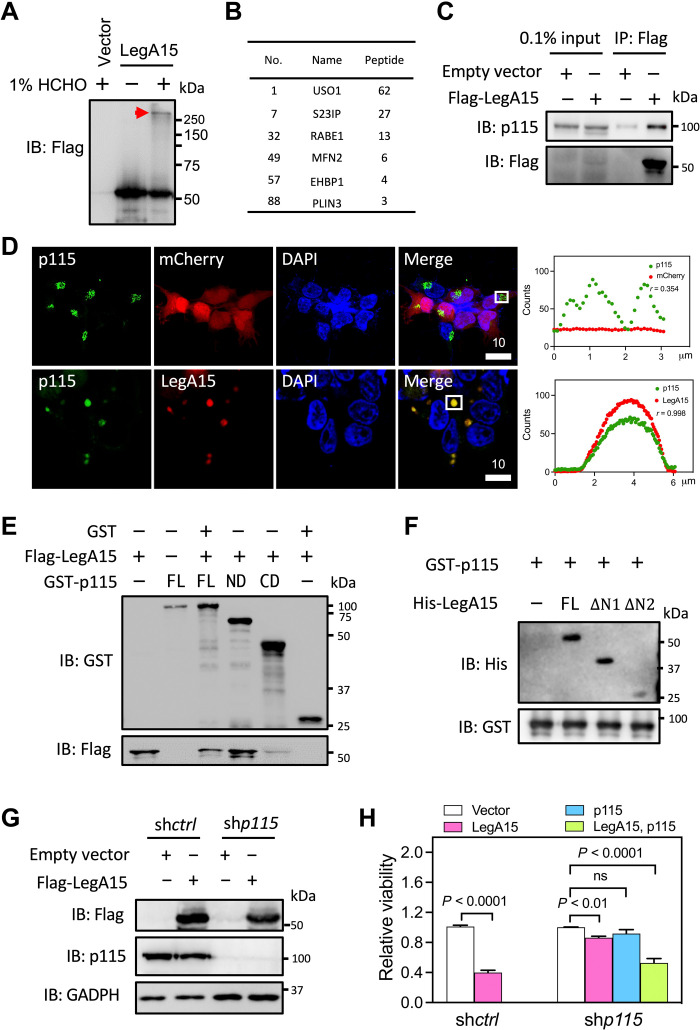
Fig. 2. LegA15 interacts with host vesicular transport factor p115.
(A) Immunoblotting (IB) analysis of the IP fraction. A cross-linked protein complex was positioned by a red arrow. (B) Putative LegA15 interaction proteins. A full list of proteins in vesicle trafficking and membrane fusion are table S1. (C) LegA15 interacts with endogenous p115. LegA15 was expressed in HEK293T and immunoprecipitated with anti-Flag antibody, and p115 was detected by anti-p115 antibody. (D) LegA15 colocalizes with p115 in the HEK293T cells expressing LegA15-mCherry. p115 in green was stained with anti-p115 antibody. Nuclei were in blue. Scale bars are in micrometer scale. Shown on the right were colocalization analyses with Pearson correlation (r) on ROI in white boxes as Fig. 1. (E) The N-terminal domain of p115 interacts with LegA15 in a GST pulldown experiment. GST-p115 fusion proteins [the full-length, ND (1 to 651) and CD (652 to 964)] were used to pull-down Flag-LegA15, and detected by anti-Flag antibody. Protein loading was examined by anti-GST antibody. (F) His-LegA15 [the full-length, ΔΝ1 (95 to 471) and ΔΝ2 (195 to 471)], GST-p115 (1 to 651) was pulled down and detected by anti-His antibody with protein loadings by anti-GST antibody. (G) p115-knockdown HEK293T cell line expressing LegA15. Flag-LegA15 was expressed for 16 hours, and detected by p115 and Flag antibodies. GAPDH, glyceraldehyde-3-phosphate dehydrogenase. (H) Knockdown of p115 suppresses the cytotoxicity of LegA15 after transfection for 36 hours. Transfection with its empty vector was a negative control. Shown is relative viability from three independent experiments and analyzed by one-way analysis of variance (ANOVA) assay. ns, not significant.

