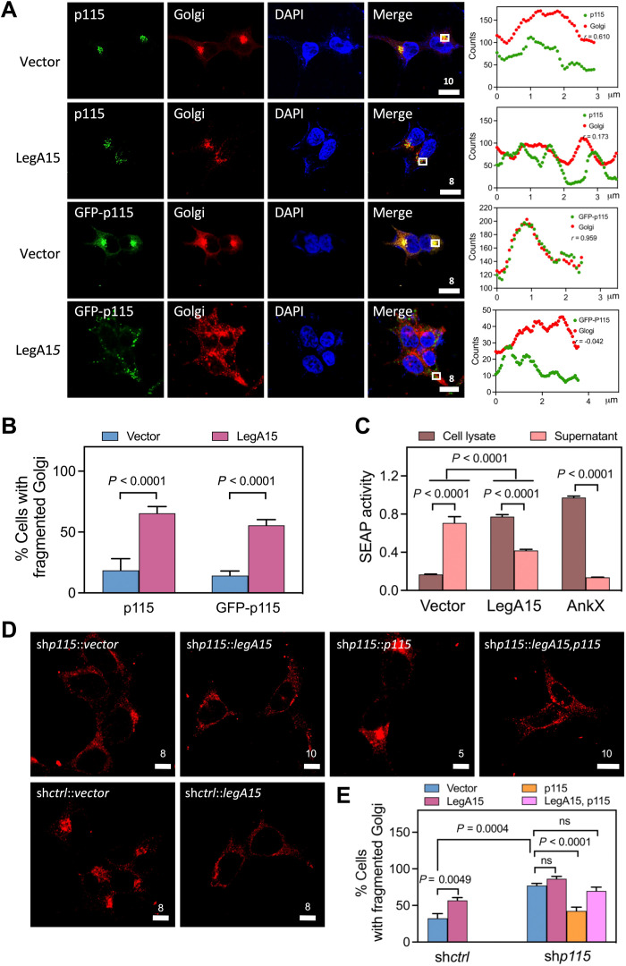
Fig. 3. LegA15 induces fragmentation of the host Golgi apparatus.
(A) Golgi apparatus localization of p115 is affected by overexpressed Flag-LegA15. GFP-p115 and endogenous p115 were detected by anti-p115 antibody in green. Golgi apparatus was stained with anti-syntaxin 6 antibody in red. Nuclei were in blue. Scale bars are in micrometer scale. Shown on the right were colocalization analyses with Pearson correlation (r) on ROI in white boxes as Fig. 1. Scale bars are in micrometer scale. (B) LegA15 induces fragmentation of Golgi apparatus in host cells. The quantification of (A) was calculated for three groups using more than 100 randomly selected cells as each group. The significance was calculated by t test. (C) LegA15 inhibits the secretion of SEAP. The HEK293T cells coexpressed with Flag-LegA15 and SEAP for 16 hours were used to measure the SEAP activity in culture medium and cell lysates, separately. Transfection with its empty vector was used as a negative control. In addition, Legionella effector AnkX was used as a positive control. Quantitation shown was from three independent experiments and analyzed by one-way ANOVA assay. (D and E) The host p115 is required for LegA15 induced Golgi fragmentation. LegA15 was overexpressed in the HEK293T cells with p115 knockdown and complemented p115. The Golgi apparatus was stained with anti-syntaxin 6 antibody (red). Nuclei were in blue. Transfection with an empty vector was a negative control. The quantification of Golgi fragmentation (E) was calculated for three groups using more than 100 randomly selected cells as one group. The significance was calculated by t test.

