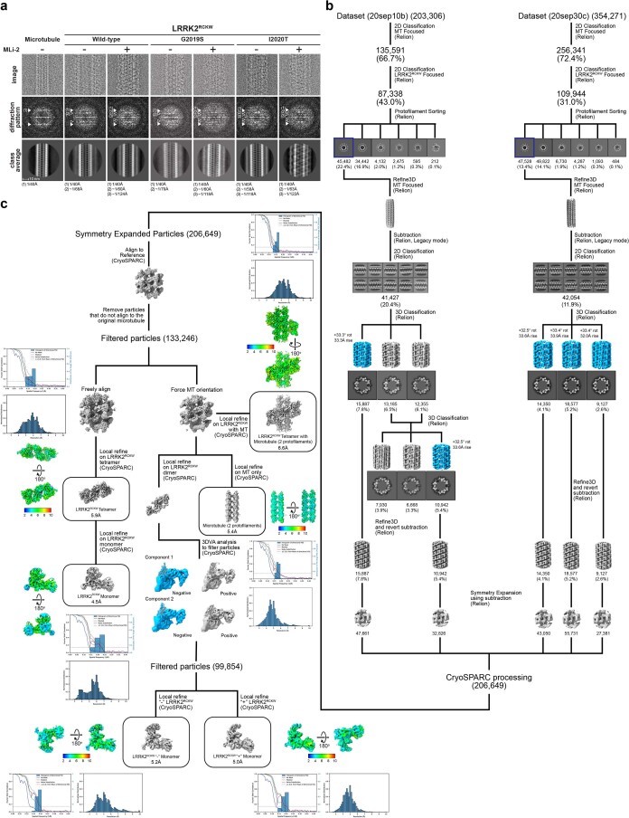
Extended Data Fig. 1. Cryo-EM structure determination of microtubule-associated filaments of LRRK2RCKW[I2020T] in the presence of MLi-2.
a, Optimization of in vitro reconstituted microtubule-associated LRRK2RCKW filaments. Top row, cryo-EM images of an individual microtubule (left) or individual microtubule-associated LRRK2RCKW filaments. Middle, Diffraction patterns calculated from the images above. Arrowheads point to layer lines arising from the microtubule (white) or from the LRRK2RCKW filaments (open), and their frequencies are indicated below the images. Bottom, 2D class averages from multiple images equivalent to those shown at the top. The type of LRRK2RCKW (WT, G2019S, or I2020T) and the presence or absence of MLi-2 during filament reconstitution are indicated at the top. b, Schematic of data processing pipeline used to obtain the different reconstructions of the microtubule-associated LRRK2RCKW[I2020T] filaments in the presence of MLi-2 (see Methods for details). Local resolution maps, Fourier Shell Correlation, directional FSC plots, and the distribution of voxel resolutions are shown for all reconstructions discussed in the text.

