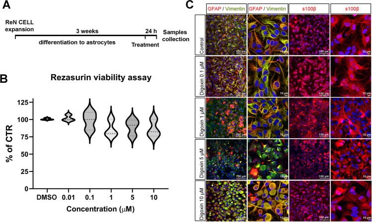
Fig. 1.
Characterization of Ren-derived astrocytes after digoxin exposure. A Diagram of the culture and treatment procedure. B Resazurin viability assay for astrocytes exposed to different concentrations of digoxin for 24 h. Results are displayed as violin with data points, for each group n = 7 samples obtained in 2 independent experiments. Statistical analysis for the effects of digoxin was performed using the non-parametric Kruskal–Wallis test followed by Dunn’s multiple comparison test. C Confocal images of immunostaining for GFAP, vimentin, and S100B a nuclei are stained with Hoechst (blue)

