. 2022 Dec 15;23(12):1014–1027. doi: 10.1631/jzus.B2200346

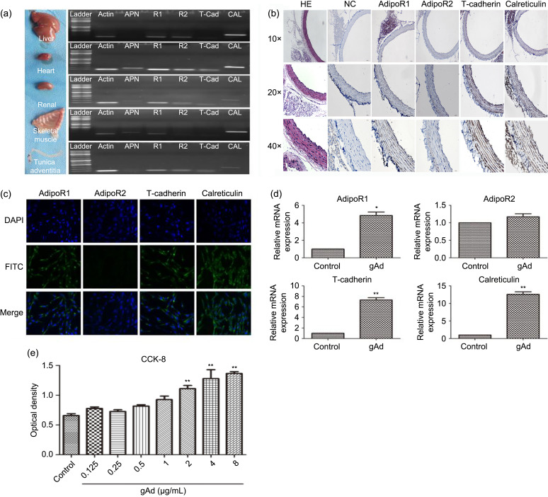
Fig. 4. Expression of adiponectin and its receptors in different organs, vessels, and AFs. (a) Adiponectin, adipoR1, adipoR2, T-cadherin, and calreticulin mRNA expression in liver, heart, renal, skeletal muscle, and tunica adventitia of vessels. Representative data from three independent experiments are shown. (b) Distribution of adipoRs in rat thoracic aorta. (c) Immunofluorescence staining in AFs revealed that the fibroblast cell membrane was the main location of adipoRs. Plasmalemma localization was revealed using FITC, and nuclear localization was revealed with DAPI. Magnification 200×. (d) The mRNA expression levels of the adipoR1, adipoR2, T-cadherin, and calreticulin proteins in AFs were investigated by qPCR. (e) AFs were grown into a 96-hole cell culture plate. After AFs were stimulated with different concentrations of gAd (0, 0.125, 0.25, 0.5, 1, 2, 4, and 8 μg/mL), proliferation of AFs was evaluated by CCK-8 using a 450-nm wavelength for optical density measurements. A concentration-dependent effect of gAd on AF proliferation was observed. The results are shown as mean±standard error of the mean (SEM), n=3. * P<0.05, ** P<0.01 vs. control. AFs: adventitial fibroblasts; AdipoR: adiponectin receptor; mRNA: messenger RNA; FITC: fluorescein isothiocyanate; DAPI: 4',6-diamidino-2-phenylindole; qPCR: quantitative real-time polymerase chain reaction; CCK-8: cell counting kit-8; gAd: globular adiponectin; HE: hematoxylin-eosin; NC: negative control.

Fig. 4