. 2022 Dec 15;23(12):1014–1027. doi: 10.1631/jzus.B2200346

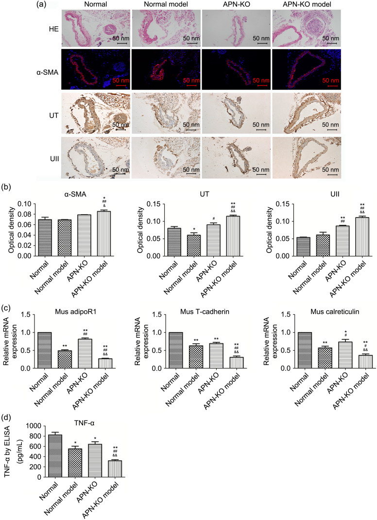
Fig. 7. Changes of α-SMA and TNF-α in the adventitial inflammation model. (a) HE staining showed the structure of each layer of the common carotid artery in the inflammation model. α-SMA expression in common carotid adventitia was examined by immunofluorescence, while UT and UII expression was examined by immunohistochemistry. Scale bar=50 nm. (b) Optical density of the target proteins in the inflammation model (IPP6.0 software was used to analyze the optical density of immunofluorescence and immunohistochemistry sections). (c) Vascular tissue was taken from the modeled part and RNA was extracted. The mRNA expression of adipoR1, T-cadherin, and calreticulin was examined by qPCR analysis. (d) TNF-α changes in mouse plasma in the inflammation model were tested by ELISA. Data are expressed as mean±standard error (SE), n=3. * P<0.05, ** P<0.01 vs. normal group; # P<0.05, ## P<0.01 vs. normal model group; & P<0.05, && P<0.01 vs. APN-KO group. α-SMA: α-smooth muscle actin; TNF-‍‍α: tumor necrosis factor-α; HE: hematoxylin-eosin; UII: urotensin II; UT: UII receptor; mRNA: messenger RNA; AdipoR: adiponectin receptor; qPCR: quantitative real-time polymerase chain reaction; ELISA: enzyme-linked immunosorbent assay; APN-KO: adiponectin-knockout; Mus: mouse.

Fig. 7