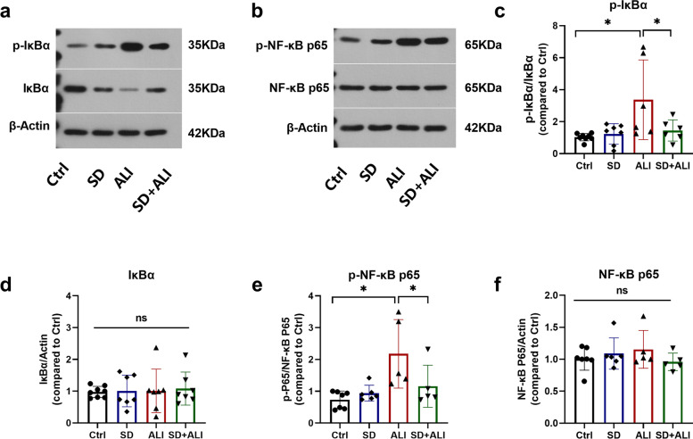
Fig. 3.
SD inhibiting ALI mice lung NF-κB signaling. a Sample western blot of p-IκBα, IκBα, and β-Actin proteins in 4 groups of mice lung(n = 5–8); b Sample western blot of p- NF-κB p65, NF-κB p65 and β-Actin proteins in 4 groups of mice lung(n = 5–8); Groups protein level data analysis of p-IκBα (c), IκBα (d), p-NF-κB p65 (e), NF-κB p65 (f), bands were semi-quantitatively analyzed by using the ImageJ software and normalized to β-actin. Data represent the mean ± sd. *p < 0.05; **p < 0.01; ***p < 0.001; NS, no statistically significant difference. The samples were derived from the same experiment and the gels/blots were processed in parallel. All of the full-length blots/gels or membrane edges visible blots are presented in Additional file 2

