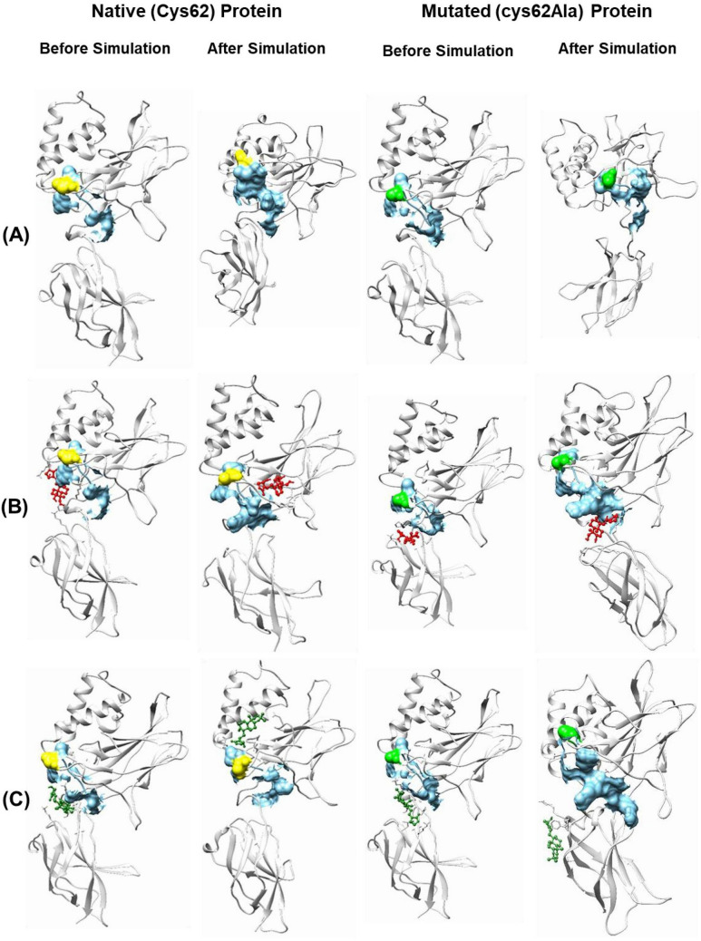
Fig. 6.
Representation of the binding site of NF-κB p50 protein (both native and mutated) and its interaction with AGP and Ana2 before and after the simulation. A Protein, B AGP, and C Ana2. Color code: light grey = protein, sky blue surface = binding site, yellow = Cys62 amino acid residue, green = Ala62 amino acid residue, red ball and stick model = AGP, and dark green ball and stick model = Ana2

