
Fig. 3.
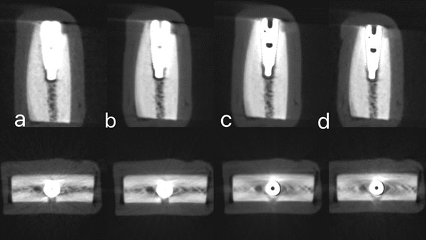
Sagittal and axial images with simulated peri-implant dehiscence defect obtanied with different four radiographic protocols. a High-dose CBCT without MAR. b Low-dose CBCT without MAR. c High-dose with MAR. d Low-dose CBCT with MAR
Official websites use .gov
A
.gov website belongs to an official
government organization in the United States.
Secure .gov websites use HTTPS
A lock (
) or https:// means you've safely
connected to the .gov website. Share sensitive
information only on official, secure websites.
Sagittal and axial images with simulated peri-implant dehiscence defect obtanied with different four radiographic protocols. a High-dose CBCT without MAR. b Low-dose CBCT without MAR. c High-dose with MAR. d Low-dose CBCT with MAR