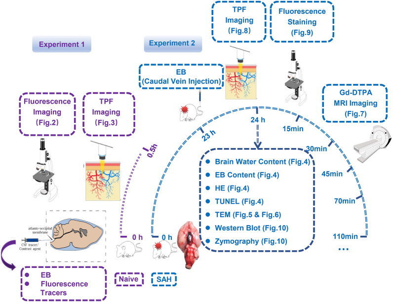
Fig. 1.
The protocol of the study. In Experiment 1, the fluorescence tracers with different molecular weights and EB dye were injected into the cisterna magna of naive rats, and at 0.5 h after injection, the individual distribution patterns of these fluorescence tracers were explored using TPF imaging. The distribution pattern of EB was observed before and after dcLN ligation. In Experiment 2, the pattern of IPAD was analysed at 24 h after establishment of SAH model. TPF imaging was applied to explore the distribution changes of these fluorescence tracers, and the ISF flow alteration after SAH was analyzed using Gd-DTPA MRI technique. The disruption of BBB, apoptosis of endothelial cells and basement membrane degradation were revealed using EB fluorescence imaging, TUNEL, MMP9 and IV-Col staining and MMP9 zymography. Three rats were randomly selected in each group involved in each item of experiment 1, and 5 rats were randomly selected in each group involved in each item of experiment 2. Abbreviations: dcLN, deep cervical lymph node; EB, Evans Blue; Gd-DTPA, gadolinium-diethylenetriaminepentaacetic acid; IPAD, intramural periarterial drainage; ISF, interstitial fluid; IV-Col, collage type IV; MMP9, matrix metalloproteinase 9; MRI, magnetic resonance imaging; SAH, subarachnoid hemorrhage; TEM, transmission electron microscopy; TPF, two-photon fluorescence; TUNEL, TdT-mediated dUTP Nick-End Labeling; WB, western blot

