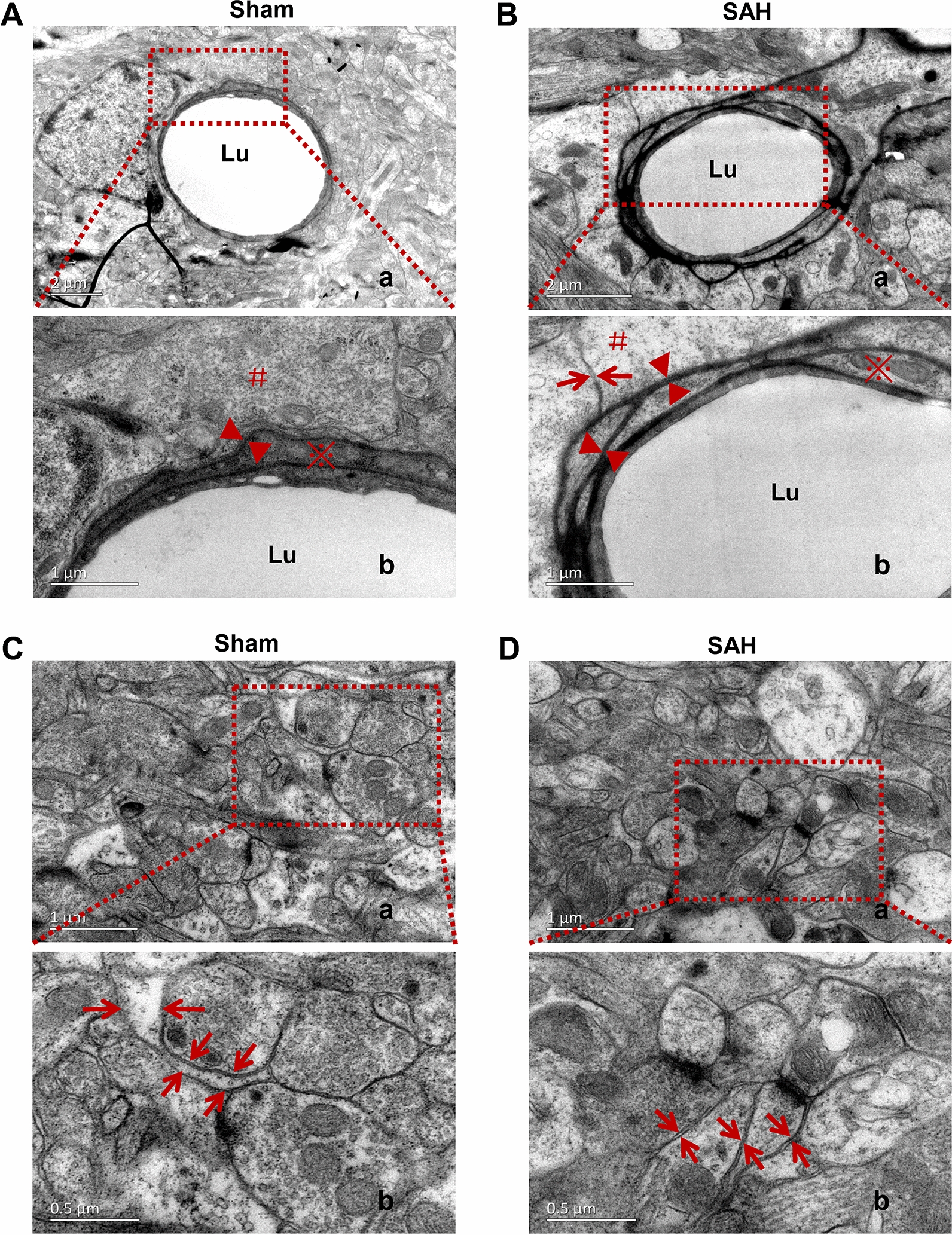
Fig. 6.

Narrowing of the ECS following IPAD injury after SAH. Compared with that of Sham group (A), the width of the IPAD pathway was significantly increased after SAH (B), suggesting IPAD impairment. The ECS around the capillaries in the frontal cortex were markedly narrowed after SAH due to the defects in the basement membranes and astrocyte swelling (A, B, C, D). n = 5 per group. A.b, B.b, C.b and D.b are enlarged images of the framed areas in A.a, B.a, C.a and D.a, respectively. “Arrow” indicates ECS; “▲” indicates the IPAD; “#” indicates the endfoot of astrocyte; “※” shows the endothelial cells. A.a, B.a: scale bar = 2 μm; A.b, B.b, C.a, D.a: scale bar = 1 μm; C.b, D.b: scale bar = 0.5 μm. Abbreviations: ECS: extracellular space; IPAD: intramural periarterial drainage; SAH: subarachnoid hemorrhage; Lu: microvessel lumen
