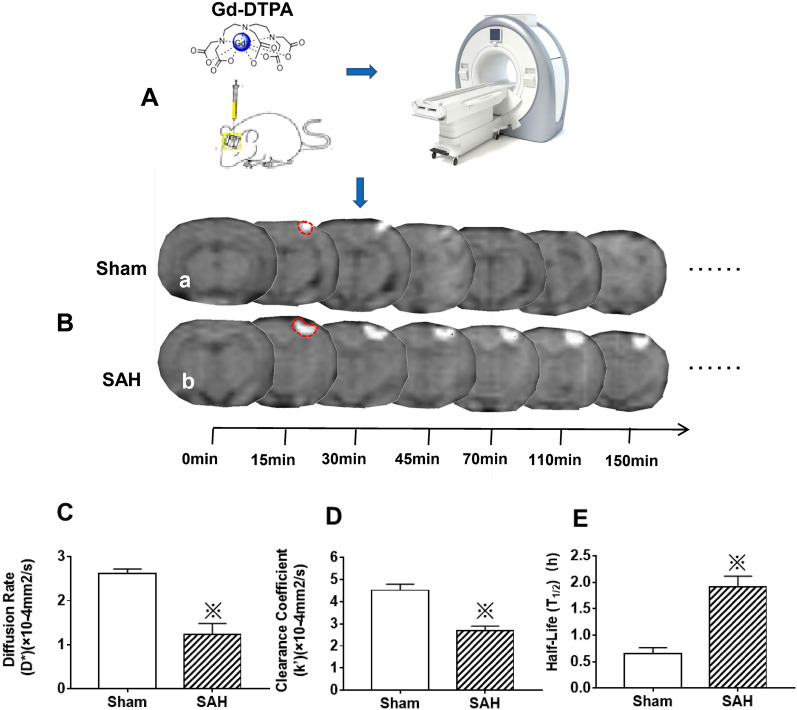
Fig. 7.
The ISF flow injury detected by MRI tracking technique after SAH. Following Gd-DTPA injection (A), the diffusion rate (D*), clearance rate (k') and the half time of Gd-DTPA were measured at 0, 15, 30, 45, 70, 110, 150 min. The results indicated that, the signal of Gd-DTPA in cortex was gradually weakened, and in the Sham group, the Gd-DTPA was completely cleared from brain at 110 min following injection; however, the signal intensity of Gd-DTPA was still strong at 150 min in the SAH group (B). After SAH, the Gd-DTPA diffusion rate (D*) and clearance rate (k') were significantly decreased compared with those of Sham group: consequently, the half time(T1/2) of Gd-DTPA signal intensity was markedly extended (C, D, E). These results showed that there was impairment of ECS spatial conformation and ISF drainage in the ECS after SAH. “※” in C, D and E indicate P < 0.05 compared with that of Sham group, n = 5 per group. Abbreviations: ECS: extracellular space; Gd-DTPA: gadolinium-diethylenetriaminepentaacetic acid; IPAD: intramural periarterial drainage; ISF: interstitial fluid; MRI: magnetic resonance imaging; SAH: subarachnoid hemorrhage

