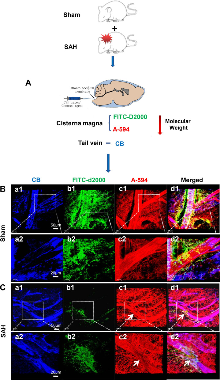
Fig. 8.
The impairment of IPAD drainage of the fluorenscence dyes. At 24 h after SAH, the FITC-D2000 and A-594 dyes were injected into cisterna magna, and the CB dye was injected into tail veins to show the microvessels in the cortex, n = 5 per group (A). At 1 h following injection, in the frontal cortex, both FITC-D2000 and A-594 flowed into the ECS and their clearance rates through IPAD were both markedly decreased compared with those of Sham group (B.b1, b2, c1, c2, C. b1, b2, c1, c2). The astrocytes surrounding the arterioles had thickened and distorted end-feet (C). B.a2-d2 are the magnified images of the framed areas in B.a1–d1; C.a2–d2 are the magnified images of the framed areas in C.a1-d1. “Arrow” in C.c1–d1, c2–d2 shows vasospasm in the arterioles after SAH. B.a1–d1, C.a1–d1: scale bar = 50 μm; B.a2–d2, C.a2–d2: scale bar = 20 μm. Abbreviations: A-594: ALEXA-594 hydrazide; CB, Cascade Blue; EB: Evans Blue; ECS: extracellular space; FITC-D2000: FITC-dextran 2000; IPAD: intramural periarterial drainage; SAH: subarachnoid hemorrhage

