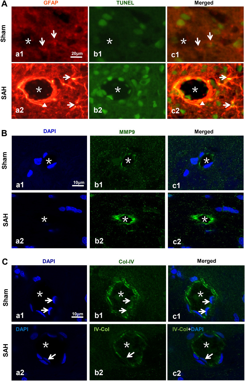
Fig. 9.
The molecules involved in IPAD impairment after SAH. In the Sham group, the astrocytes were evenly distributed around the microvessels, and there were rare apoptotic astrocytes in cortex (A.a1–c1). After SAH, the expression level of GFAP was markedly enhanced, and there were numerous apoptotic astrocytes around the microvessels (A.a2–c2). The number of astrocyte processes was increased after SAH (A.a1, a2, c1, c2). Immunofluorescence staining showed that after SAH, the endothelial MMP9 of microvessels increased significantly (B.a1–c1, a2–c2), while Col-IV decreased significantly (C.a1–c1, a2–c2). The nuclei were labeled with DAPI (B.a1, a2, C.a1, a2). n = 5 per group. “Arrow” in A shows the astrocyte; “*” indicates the microvessel lumen; “▲” indicates the microvessels; “Arrow” in C showed the nucleus. A: scale bar = 20 μm; B, C: scale bar = 10 μm. Abbreviaitons: DAP: 4',6- diamidino-2- phenylindole; Col-IV: Collagen Type IV; GFAP: glial fibrillary acidic protein; IPAD: intramural periarterial drainage; MMP9: Matrix metalloproteinase 9; SAH: subarachnoid hemorrhage

