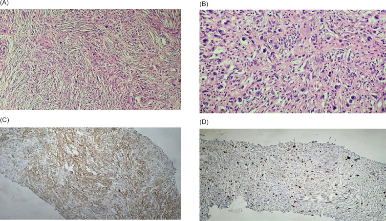
FIGURE 2:
(A) Short fascicles of spindled myofibroblastic cells without overt atypia admixed with acute and chronic inflammatory cells (200×). (B) Ganglion-like polygonal cells with large rounded nuclei and prominent nucleoli (400×). Immunohistochemistry (IHC) analyses revealed positive reaction for (C) smooth muscle actin (SMA) and (D) Ki-67 (5%).

