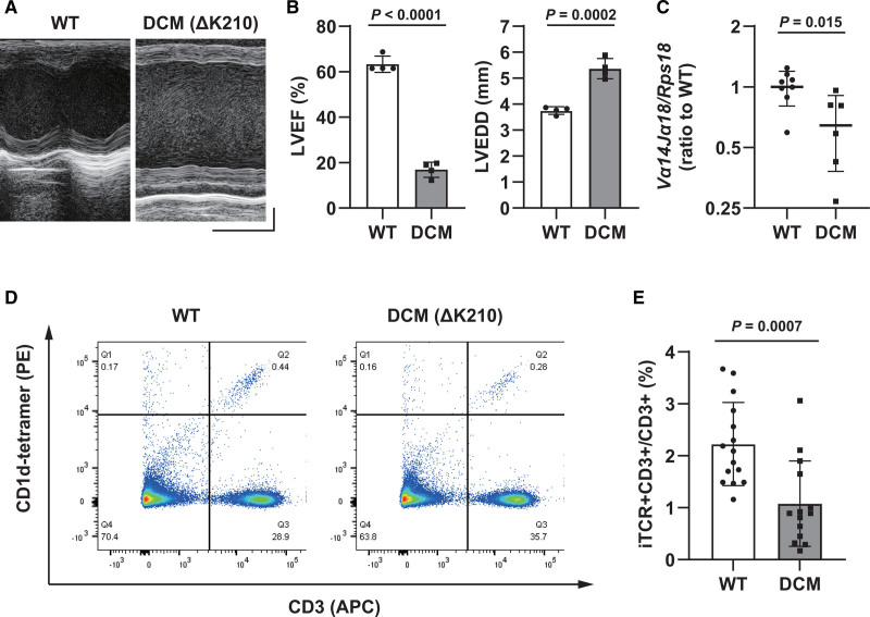
Figure 2.
Characterization of dilated cardiomyopathy (DCM) mice (5 weeks old). A, Representative echocardiographic images of wild-type (WT) and DCM mice. Horizontal scale indicates 100 ms‚ and vertical scale indicates 1 mm. B, Left ventricular ejection fraction (LVEF, left) and left ventricular end-diastolic diameter (LVEDD, right) in WT and DCM mice (n=4). C, Gene expression of invariant T cell receptor (TCR; Vα14Jα18) in the myocardium of WT and DCM mice, measured using reverse transcription-quantitative polymerase chain reaction (n=8 and 6, respectively). D, Flow cytometry data of the spleens of WT and DCM mice. CD1d-tetramer binding (PE)- and CD3 (allophycocyanin [APC])-positive cells were defined as invariant natural killer T (iNKT) cells. E, Quantification of the percentage of iNKT cells to CD3-positive cells (n=15 and 14, respectively). Data are presented as mean±SD. Statistical significance was determined using Student t test.

