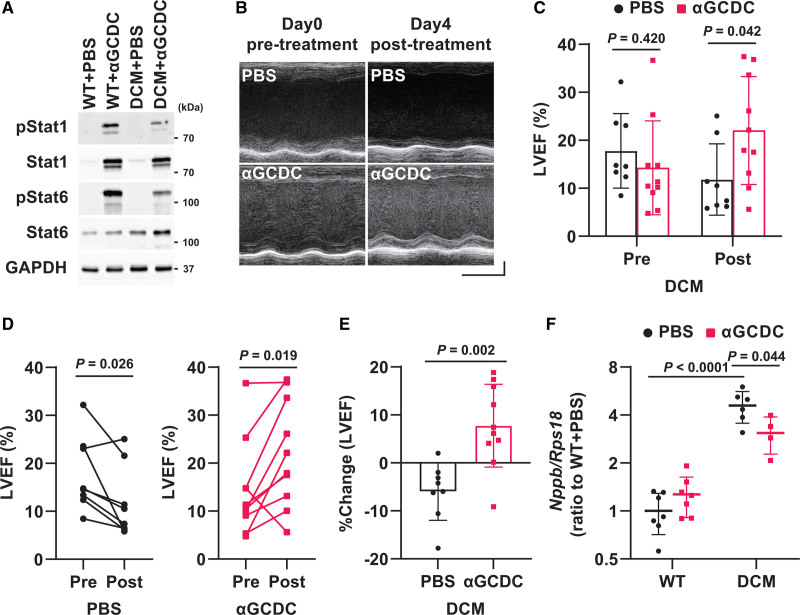
Figure 4.
Acute effect of α-galactosylceramide (αGalCer)-pulsed dendritic cells (αGCDCs) on the cardiac function of dilated cardiomyopathy (DCM) in mice. A, Phosphorylation of Stat1 and Stat6 in the myocardium on day 4 following αGCDC treatment. Glyceraldehyde-3-phosphate dehydrogenase (GAPDH) was used as a loading control. B, Representative echocardiographic images of DCM mice at pretreatment and posttreatment (day 4) with PBS and αGCDCs. Horizontal scale indicates 100 ms, and vertical scale indicates 1 mm. C, Left ventricular ejection fraction (LVEF) in DCM mice at pretreatment (Pre) and posttreatment (Post; day 4) with αGCDCs (n=8–10). D, Individual changes in LVEF; DCM+PBS, n=8; DCM+αGCDC, n=10. E, Percent change in the LVEF from the beginning (pretreatment) to the end of the study (day 4 following each treatment, posttreatment) in mice treated with PBS and αGCDCs (n=8–10). F, Gene expression of BNP (brain natriuretic peptide, Nppb) in the myocardium of wild-type (WT) and DCM mice on day 4 after αGCDC treatment, measured using reverse transcription-quantitative polymerase chain reaction (n=4–7). Data are presented as mean±SD. Statistical significance was determined using Student t test for C and E, paired Student t test for D, and 1-way ANOVA with a post hoc test (Tukey) for F.

