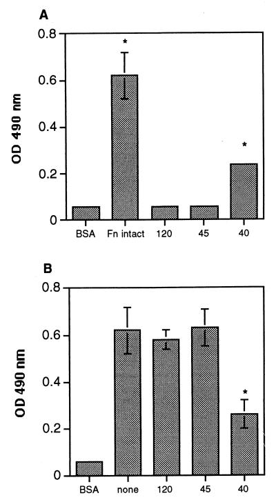
FIG. 4.
Adhesion of A. fumigatus conidia to fibronectin protein fragments. Microtiter plates were coated with intact fibronectin (Fn intact) or fibronectin fragments containing the gelatin-binding domain (45 kDa [45]), the cell binding domain (120 kDa [120]), or the GAG binding domain (40 kDa [40]) and tested for the ability to promote spore adhesion. Background wells contained BSA only. Peroxidase-labeled conidia were added to wells containing fragments alone (A) or were preincubated with the fragments before conidia were added to wells coated with fibronectin (B). Unbound conidia were removed by washing the plates with PBS–0.05% Tween 20, and the number of bound conidia was determined by measuring the optical density (OD) at 490 nm. The values are the means ± standard deviations of triplicate wells, and the figures are representative of two independent experiments. ∗, P < 0.05 versus BSA (for panel A) or versus none (for panel B).

