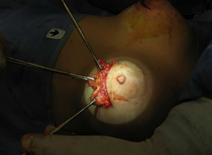
Fig. 1.
Intraoperative view of the subareolar superior pedicled glandular flap that it is harvested from the upper pole of the areola, where bulging is evident; then, it is transferred to the lower pole. The flap is elevated through an “inverted V” glandular incision performed in the upper pole of areola where nipple-areola complex bulging is more evident. Following this, we then split the distal portion of the glandular flap into three or four little tongues.

