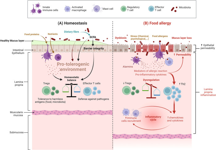
Figure 1.
Intestinal barrier in a steady vs. food allergy state. (A) Under homeostatic conditions (cohesive intestinal barrier, diverse and active microbiota, exposure to food antigens), antigen-presenting cells promote the induction of food antigen-specific Treg cells. These cells induce tolerance to dietary antigens by a range of mechanisms, including inhibition of antigen-specific T helper type 2 (Th2) cell responses, suppression of pathogenic Th2 cell-like reprogramming of T cells and of mast cell activation, and the production of barrier-protective cytokines; SCFA: short-chain fatty acids (B) In food allergy, dysbiosis associated with an impaired gut barrier compromise the differentiation of naive T cells into Treg cells and instead leads to the differentiation of type 2 adaptive T helper cells (Th2) and inflammation – more details are provided in the text and in Figure 2 (Created with BioRender.com).

