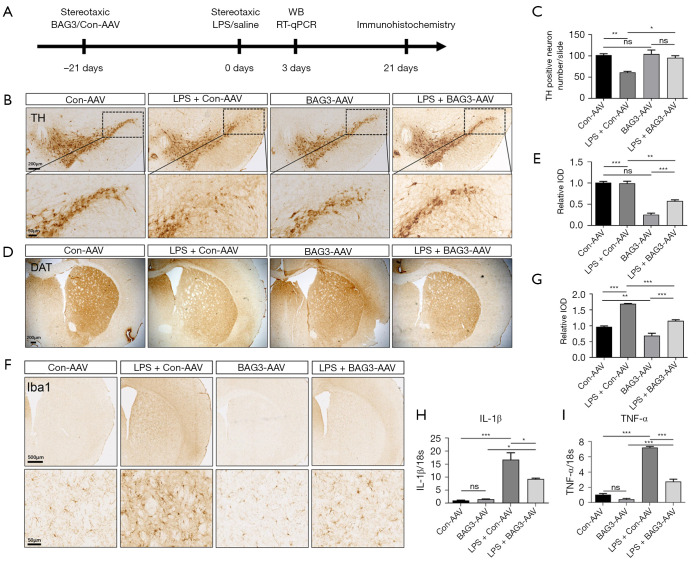
Figure 4.
Overexpression of BAG3 reduced LPS-induced TH loss and microglial activation. (A) Experimental flow chart. BAG3-AAV or Con-AAV was injected into the bilateral striatum of C57BL/6 mice. After 21 days, LPS or PBS was injected into the bilateral striatum. (B,C) At 21 days after LPS injection, immunostaining and quantification of TH-positive neurons in mice. (D,E) At 21 days after LPS injection, immunostaining and quantification of DAT in mice. (F,G) At 21 days after LPS injection, immunostaining and quantification of Iba1+ cells in mice. (H,I) At 3 days after LPS injection, expression of IL-1β and TNF-α was demonstrated by RT-qPCR and in statistical histograms. n=3; mean ± SEM; *P<0.05, **P<0.01, ***P<0.001; ns, not significant. BAG3, Bcl2-associated athanogene 3; AAV, adeno-associated virus; LPS, lipopolysaccharide; WB, Western blotting; RT-qPCR, reverse transcription quantitative polymerase chain reaction; TH, tyrosine hydroxylase; DAT, dopamine transporter; IOD, integrated option density; TNF, tumor necrosis factor; IL, interleukin; SEM, standard error of mean.

