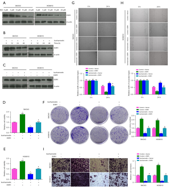
Figure 6.
ISO inhibits proliferation, migration and invasion of OC cells by targeting ESR1. (A) ESR1 protein levels in SKOV3 and HO8910 cells treated with ISO (0–20 µM) for 24 h. (B) Protein levels of ESR1 in SKOV3 and HO8910 cells treated with 15 µM ISO for different lengths of time. (C) ESR1 protein levels in SKOV3 and HO8910 cells after ESR1 transfection and 15 µM ISO treatment alone or in combination. (D,E) Cell viability. (F) Colony formation. Crystal violet staining; magnification, ×1. (G,H) Wound healing and images of monolayers were captured under an optical light microscope. Scale bar =200 µm. (I) Cell invasion. Crystal violet staining; scale bar =200 µm. Compared with the Control group, *, P<0.05; compared with the ISO group, #, P<0.05. ISO, isorhamnetin; OC, ovarian cancer; ESR1, estrogen receptor 1.

