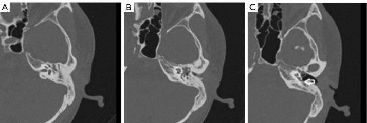
Figure 1.
CT of a patient with stage III adhesive otitis media. (A) A patient with stage III adhesive otitis media. Computed tomography suggests a soft tissue shadow of the right epitympanum and tympanic antrum, and the ossicular chain is surrounded by a soft tissue shadow. (B) The mesotympanum-hypotympanum was invaginated with a soft tissue shadow. (C) A mesotympanum-hypotympanum invagination and a tight fit with the promontory (white arrow).

