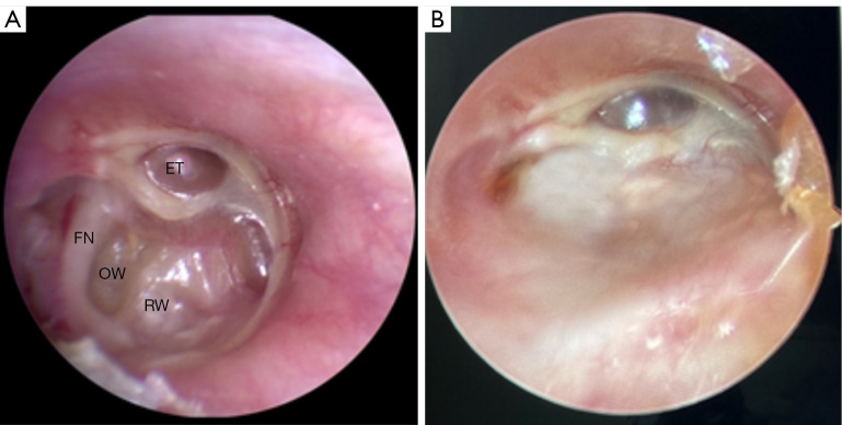
Figure 3.
Preoperative and postoperative comparison of the patient with stage IV adhesive otitis media. (A) A preoperative endoscopy of a patient with stage IV adhesive otitis media, showing that the tympanic membrane was thin and invaginated. The endoscopy was unable to visualize the full extent of the adhesions. (B) An endoscopy of a stage IV adhesive otitis media patient at 3 months after tympanoplasty type III surgery, showing good tragus cartilage-chondrogenic healing. ET, eustachian tube-tympanum opening; FN, tympanic segment of the facial nerve; OW, oval window; RW, round window.

