Fig. 8.
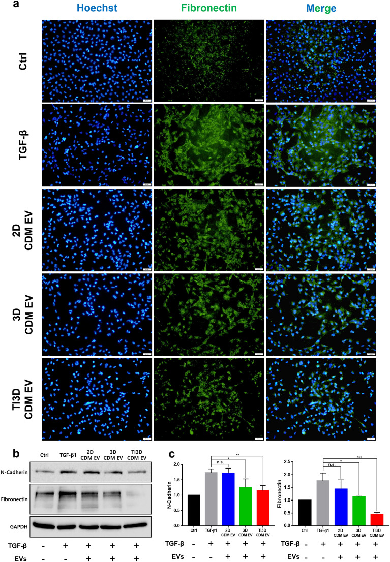
Anti-fibrotic properties of EVs. a The fluorescence-based immunocytochemistry of fibronectin expression of TGF-β induced fibrosis condition in HK2 cells. b Western blot analysis for representative fibrosis markers after EVs treatments in fibrosis induced-HK2 cells. c Quantification data for N-cadherin and fibronectin expression from Western blot analysis (analysis with Image J, values are presented as mean ± SD (n = 3) and statistical significance was obtained with one-way analysis of ANOVA with Tukey’s multiple comparison post-test (*p < 0.05; **p < 0.01; ***p < 0.001; ****p < 0.0001))

