Figure 1.
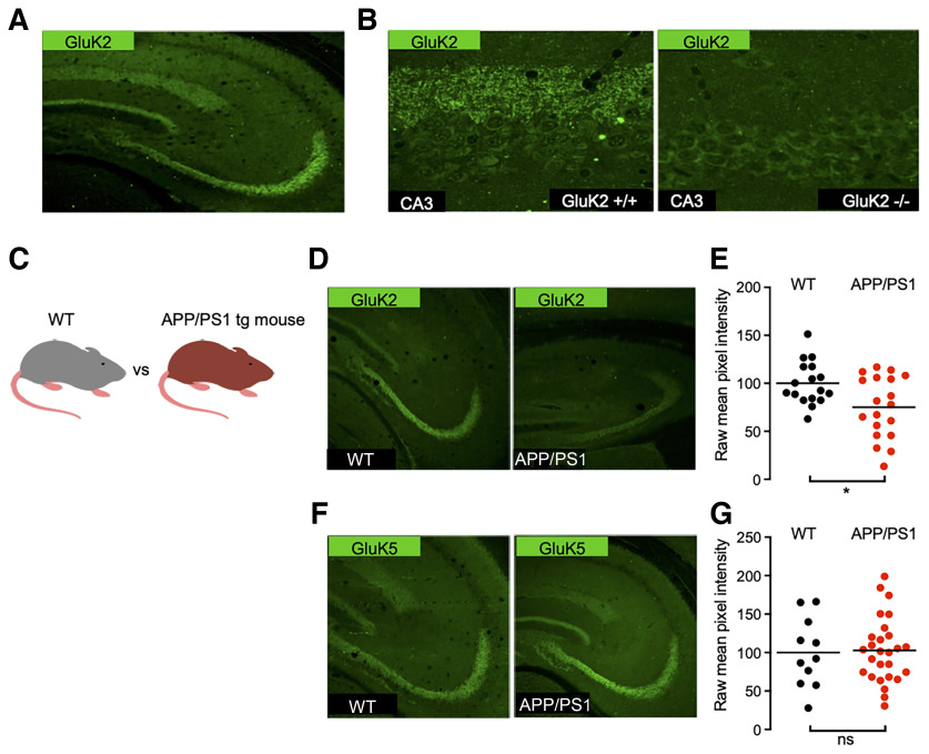
Distribution of kainate receptor subunits GluK2 and GluK5 in the hippocampus of WT and APP/PS1 mice. A, Immunolabeling of mouse hippocampal sections reveals strong expression of GluK2 subunit in the stratum lucidum. B, Zoom-in acquisitions on the CA3 area show staining adjacent to the CA3 pyramidal layer in the stratum lucidum corresponding to Mf–CA3 synapses. This staining is lost in GluK2−/− mice. C, Cartoon representing an APP/PS1 mouse model compared with a WT littermate. D, GluK2 staining in the CA3 region of APP/PS1 mice is decreased in comparison to WT. E, Quantification of raw mean pixel intensity in the stratum lucidum region of images as in D. Data were normalized to the mean intensity of the WT condition. F, GluK5 staining in the CA3 region of the APP/PS1 mice is similar to that for WT mice. G, Quantification, as in E, shows no detectable decrease of GluK5. *p < 0.05 and ns = non-significant.

