Figure 4.
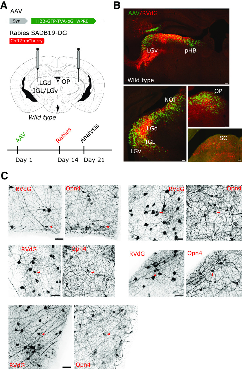
Retrograde transsynaptic labeling of the retina independent of the genetic identity of the starter cells. A, Scheme of the delivery strategy of a constitutive AAV and monosynaptic restricted rabies to the retinorecipient regions of the prethalamus, thalamus, pretectum, and midbrain, showing the timeline of viral injections. B, Infection with the constitutively active helper AAV can be detected by its expression of nuclear-restricted GFP (green) in several retinorecipient areas of the diencephalon and midbrain. Primary infection with a RVdG-mCherry2 virus (red) is detected abundantly in the retinorecipient LGd, IGL, pHB, OP and to a lesser extent in the LGv and SC. Scale bars: 100 µm. C, Transsynaptic spread of the RVdG to the retinas labels large numbers and types of RGCs, including those with high melanopsin (Opn4) expression (arrowheads). Images are a representative example from one tracing experiment out of three with similar infection patterns. Scale bars: 50 µm.

