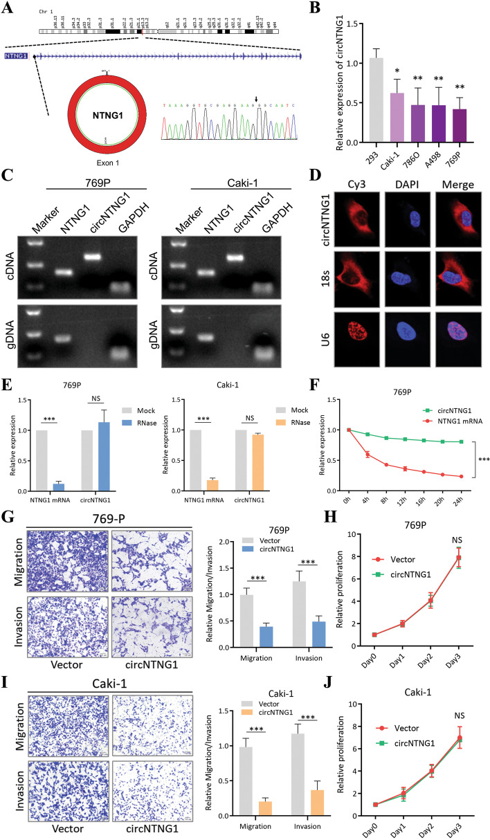
Fig. 2.
Characteristics of circNTNG1 in RCC. a. Chromosomal origin and Sanger sequencing confirmation of circNTNG1. b. Expression of circNTNG1 in normal kidney cell line 293 and four RCC cell lines (Caki-1, 786-O, A498 and 769P). c. DNA electrophoresis of circNTNG1 and linear NTNG1 from cDNA and gDNA in 769P and Caki-1. GAPDH was used as positive control. d. FISH experiment detecting the subcellular localization of circNTNG1. Eighteen s was used as positive cytoplasm control. U6 was used as positive nucleus control. e. RNase treatment assay of circNTNG1 and linear NTNG1 in 769P and Caki-1 cells. The RNA levels were determined by qRT-PCR. Expression levels were normalized to the mock group. f. Actinomycin D assays of circNTNG1 and linear NTNG1 in 769P cells. The RNA levels were determined by qRT-PCR. Expression levels were normalized to 0 h. g. Representative images (left) and quantification (right) data of Transwell migration/invasion assay of 769P cells with vector/circNTNG1-overexpression. Cell number was determined by counting five random fields under microscope. h. Proliferative activity of 769P cells with vector/circNTNG1-overexpression measured by CCK8 assay. Levels were normalized to day 0. i. Representative images (left) and quantification (right) data of Transwell migration/invasion assay of Caki-1 cells with vector/circNTNG1-overexpression. Cell number was determined by counting five random fields under microscope. j. Proliferative activity of Caki-1 cells with vector/circNTNG1-overexpression measured by CCK8 assay

